

Uterus and lining
Scans help assess fibroids, features of adenomyosis, the endometrium and the shape of the uterus. Some cavity concerns, including suspected adhesions, may require additional tests.


A closer look at your uterus, ovaries and cycle to help plan your next step.
Talk to a Fertility SpecialistFERTILITY ASSESSMENT AT SUNFLOWER
Transvaginal ultrasound (TVS) is a useful part of evaluating fertility. At Sunflower Infertility and IVF Centre, it helps specialists examine the uterus, ovaries and surrounding pelvic structures, track ovulation and make informed treatment decisions.
A fertility-focused scan looks at specific questions: how follicles are developing, how the uterine lining changes through the cycle, and whether a structural finding needs further investigation. Doppler imaging may also be used to assess blood flow.

Ultrasound can reveal findings that are not apparent from symptoms alone. Your specialist interprets these alongside your history, examination and other fertility tests.
Scans help assess fibroids, features of adenomyosis, the endometrium and the shape of the uterus. Some cavity concerns, including suspected adhesions, may require additional tests.
TVS can identify ovarian cysts, including endometriomas, polycystic ovarian appearance and some pelvic or tubal abnormalities. A normal scan does not rule out every cause of infertility or endometriosis.
Ultrasound uses sound waves rather than ionising radiation. When clinically needed, repeat scans help your specialist follow follicle growth and changes in the uterine lining.
TVS is an internal examination using a vaginal probe. The procedure should be explained before it begins; tell your care team about any concerns or discomfort.
Follicular monitoring usually involves a series of scans spaced across your cycle. This helps your specialist follow change over time, rather than relying on a single snapshot.
Ultrasound also guides procedures during fertility treatment, including egg retrieval and embryo transfer, so the specialist can see the relevant structures in real time.
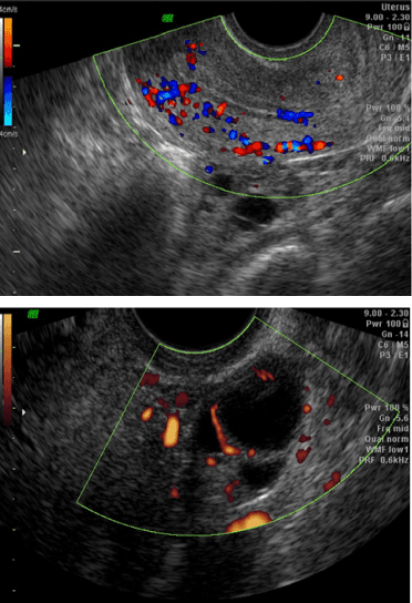
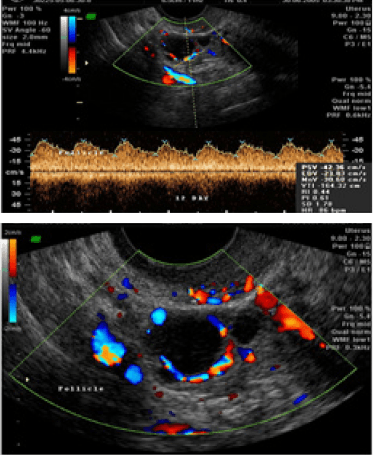
Colour Doppler adds information about blood flow to the ultrasound image. It can help assess circulation within the uterus, its lining and the ovaries.
Blood-flow findings may support follicular monitoring and the assessment of fibroids, polyps or ovarian masses. They are considered alongside the standard scan, symptoms and other results; they do not independently confirm a diagnosis or determine treatment timing.
Your specialist decides when this additional information is useful for your assessment.
A MORE COMPLETE VIEW
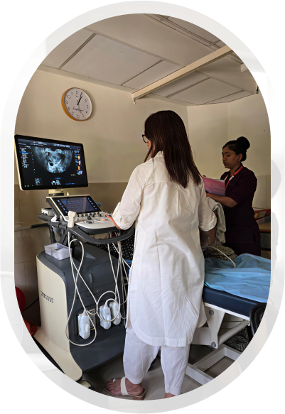
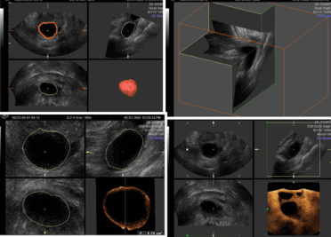
Sunflower's sonography service has five ultrasonography units with transvaginal, abdominal and linear probes, along with volume probes for 3D and 4D imaging, colour and pulsed-wave Doppler, and interventional sonography.
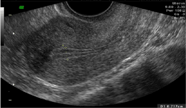
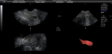
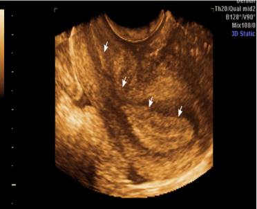
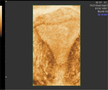
These techniques can provide additional views of the uterine cavity and help assess endometrial or follicular volume when clinically useful. Not every patient needs every imaging technique: your specialist selects the scan that answers the clinical question.
Images may show a triple-line endometrial pattern around mid-cycle, a thicker lining in the secretory phase, follicular volume before ovulation or blood flow around the endometrium and follicles.
No single image or volume measurement can establish whether a pregnancy will occur. Your specialist will explain the findings in the context of your cycle and full fertility assessment.
A fertility evaluation is generally recommended after 12 months of trying to conceive if you are under 35, or after six months if you are 35 or older. If you are over 40, an earlier assessment may be appropriate.
Seek advice sooner if you have irregular or absent periods, a known gynaecological condition or concerns about recurrent pregnancy loss. TVS may be one part of the assessment; the tests you need depend on your circumstances.
YOUR QUESTIONS
Understanding the scan can help you feel more prepared for your appointment.
It is an ultrasound examination using a transvaginal probe to look at the uterus, ovaries and nearby pelvic structures. It helps assess findings that may affect fertility and track follicular and endometrial changes through the cycle.
TVS uses sound waves and no ionising radiation. Because it is an internal examination, pressure or discomfort can occur. Tell the person performing the scan if it hurts; you can ask to pause or stop. Your care team can discuss suitability and alternatives with you.
That depends on the reason for your assessment. Follicular monitoring usually involves several visits timed around your cycle; a diagnostic assessment may need a single appointment. Your specialist will explain the schedule for you.
A routine pelvic ultrasound assesses structures at one point in time. Follicular monitoring uses a series of scans to track follicle growth, signs of ovulation and changes in the uterine lining.
It shows blood flow as well as anatomy. This can provide additional information about the uterus, endometrium, follicles or a finding that needs assessment. Your specialist interprets it alongside the rest of your results.
Not necessarily. A scan finding is one part of the overall assessment, not a prediction on its own. Your specialist will explain whether it needs further investigation and discuss the management or treatment options relevant to you.
TAKE THE NEXT STEP
Understanding your reproductive health is a starting point for a plan that fits your needs. Whether you would like to discuss an existing scan or begin a fertility assessment, our specialists can help you understand what comes next.
Talk to a Fertility Specialist