

Preimplantation Genetic Diagnosis (PGD) was developed in the late 1980s as an alternative to prenatal diagnosis and possible termination of pregnancy of an affected fetus for couples who are at risk of passing on serious genetic diseases to their children.
Preimplantation Genetic Diagnosis technique requires to test embryos for genetic disorders before it implants in the womb (uterus). It avoids the need for termination of pregnancy.
An increasing number of genetic disorders can now be diagnosed by this technique.
It should be noted that genetic disorders could be due to either a single gene disorder or more than one gene.
In cases of recurrent chromosomal abnormalities such as Down’s syndrome and recurrent miscarriages caused by parental translocations, the number and character of several chromosomes can be determined.
Controlled ovarian stimulation is done using Gonadotropins. TVS monitoring of follicles is done. Trigger is adminstered once follicles reach maturity. Egg retrieval is performed. Husband’s semen sample is obtained. ICSI is used for fertilisation. This is to reduce the risk of contamination with sperm DNA.
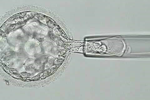
Embryo biopsy is performed on blastocysts.6-10 cells are obtained from trophoectoderm layer.The procedure is carried out under the microscope without damaging embryo’s ability to continue to develop normally (because at this stage of development none of the embryo cells have become specialized). The cell is then analyzed for the presence of genetic disorders.
Couples with genetic disorders should receive adequate counselling before embarking on PGD. They should be informed about other options such as prenatal diagnosis and termination of pregnancy if the fetus is affected, gamete donation, remain childless and adoption.
Unfortunately, for older women and women with high baseline FSH levels, PGD may not be a good alternative because not adequate embryos are formed.
It is important to note that there is a possibility that none of the embryos tested will be normal, in this case none would be suitable to transfer.
| Autosomal Recessive | Autosomal Dominant | Sex-linked | Chromosomal Abnormalities |
|---|---|---|---|
| Cystic Fibrosis | Huntingtons Disease | Hemophilia | Translocation |
| Mucopolysaccharidosis | Congenital Adrenal Hyperplasia | Duchenne Muscular Dystrophy | Klinefelter’s Syndrome |
| Thalassemia | Marfans Syndrome | Fragile X | |
| Sickle Cell Disease | Myotonic Dystrophy | X Linked Mental Retardation | |
| Spinal Muscular Dystrophy | Wiskott-Aldrich syndrome | ||
| Breast cancer (BRCA1) | |||
| Familial Adenomatous polyposis coli (FAP) | |||
| Familial Alzheimer’s disease |
Pre-implantation genetic screening (PGS), also called aneuploidy screening, involves screening embryos using NGS for 46 chromosomes. Aneuploidy is a condition where the embryo contains the wrong number of chromosomes in each cell. The doctor then selects only the chromosomally normal embryos for embryo transfer.
Advanced maternal age > 38 years
Recurrent implantation failure
Recurrent Miscarriage
Severe male factor
Controlled ovarian stimulation is done using Gonadotropins. TVS monitoring of follicles is done. Trigger is adminstered once follicles reach maturity. Egg retrieval is performed. Husband’s semen sample is obtained. ICSI is used for fertilisation. This is to reduce the risk of contamination with sperm DNA.
Embryo biopsy is performed on blastocysts.6-10 cells are obtained from trophoectoderm layer.The procedure is carried out under the microscope without damaging embryo’s ability to continue to develop normally (because at this stage of development none of the embryo cells have become specialized). The cell is then analyzed by NGS (Next Generation Sequencing) to determine chromosomal composition of each embryo that is all 23 pairs of chromosomes are analysed. embryos which are aneuploid (monosomy i.e single copy of any chromosome pair or trisomy i.e 3 copies of any chromosome or polyploid 69 chromosomes ) are discarded.
With advent of trophoectoderm biopsy and NGS turn around time is quick, results are more accurate, also cost involved is lower.
Chromosomally normal embryos are placed back into uterine cavity. This method has increased success rates.