Scrotal sonography is used to evaluate the testicle disorder and surrounding organs including prostate gland, epididymis, absent or undescended testicles and any testicle abnormality.
You need to come into the ultrasound laboratory for the test.
The test is important if a patient has acute pain in the scrotum and usually used to take images for evaluating disorders of the testicle.
If the doctor or patient found a mass in the scrotum, the scrotal sonography will help to determine whether a mass is cystic or solid. Does the mass will interfere with reproductive process or it cause any problem with normal sperm production such as tubal blockage.
It also helps to diagnose the causes of testicular pain.
It helps to determine the inflammatory condition of the testicles caused by infectious process such as a tumor that cause swelling.
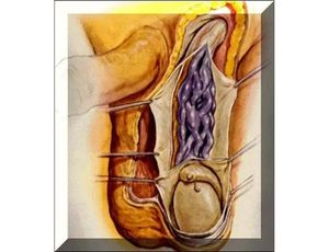
Torsion is defined as a twisting of testicle causing blocking the flow of blood to or from testicle, abnormal attachment of the epididymis to the wall of the scrotum testicle.
It can review the causes of trauma such as a hematocele (a collection of blood that surrounds the testicle), bruise or rupture of the testicle.
Sonography of the Scrotum
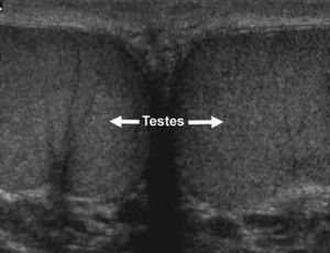
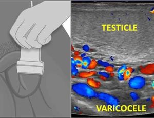
Ultrasonography (US) with a high-frequency (7.5-10-MHz) transducer has become the imaging modality of choice for examination of the scrotum. US examination can provide information valuable for the differential diagnosis of a variety of disease processes involving the scrotum that have similar clinical manifestations (eg, pain, swelling, or presence of mass). The pathologic condition that may be at the origin of such symptoms can vary from testicular torsion to infection to malignancy. The ability of color and power Doppler US to demonstrate testicular perfusion aids in reaching a specific diagnosis in patients with acute scrotal pain. This review covers the anatomy of the scrotum and the scanning protocol for scrotal US, as well as detailed descriptions of disease processes and their US appearances. Newly described conditions such as intratesticular varicoceles and other benign intratesticular cystic lesions are also discussed.
Ultrasonography (US) performed with a high-frequency transducer and the use of pulsed and color Doppler modes is the imaging modality of choice for evaluating acute and nonacute scrotal disease. Many of these disease processes, including testicular torsion, epididymo-orchitis, and intratesticular tumor, produce the common symptom of pain at presentation, and differentiation of these conditions and disorders is important for determining the appropriate treatment. US with a high-frequency transducer helps to better characterize intrascrotal lesions, and in many instances the findings suggest more specific diagnoses. High-frequency US in its present state can help identify certain benign intratesticular lesions, resulting in testes-sparing surgery. Familiarity with US characteristics and the examination pitfalls of scrotal US is essential for establishing the correct diagnosis and initiating treatment.
This review is organized on an organ basis and proceeds from superficial to deep structures. We review the anatomy of the scrotum and its contents, US scanning techniques, and US features of various pathologic conditions. This review is intended to bring the reader up to date with new technology and to provide insights into the US diagnosis of scrotal disorders. Newly described entities, such as intratesticular varicocele and other benign intratesticular cystic lesions, are discussed in detail.