Our infertility department, like obstetrics is also incomplete without the valuable aid of USG. Diagnosis of difficult and complicated gynaec pathologies which lead to infertility like endometriosis, adenomyosis, chocolate cysts, fibromyomas, hydrosalpinx & Tubo-Ovarian mass, poly cystic ovaries and ovarian cysts, Pelvic Inflammatory disease, intrauterine and intra pelvic adhesions, Septate uterus, Asherman’s syndrome, pelvic abscess and Ectopic pregnancy is possible with great accuracy and certainty by ultrasonography.
It is actually a whole lot of valuable, crucial information which the infertility and IVF specialist can get out of the follicular scans which are done by USG. Ovarian volume, follicular volume and size, peri follicular flow, endometrial thickness and pattern, myometrial echotexture, spiral artery blood flow, uterine artery blood flow and colour doppler; all the doppler indices like Resistance Index, Pulsatility index, Systolic/Diastolic ratio give us an idea of blood flow to endometrium and ovarian follicles. Ovum pick up and embryo transfer is also procedures done exclusively under USG guidance by the IVF specialist.
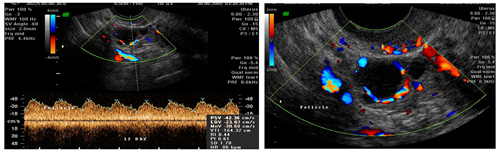
Uterine arteries supply blood to the uterus (from the internal iliac) they further divide into radial, arcuate, spiral arteries. By colour Doppler flow, we are able to assess the exact blood flow to the uterus, myometrium and endometrium. The various Doppler indices like Resistance index (RI), Pulsatility index, peak systolic velocity (PSV), end diastolic velocity (EDV), S/D ratio, gives us an idea of blood flow to endometrium and ovarian follicles. Mature follicles have high vascularity which can help in timing of hCG injection by the IVF specialist.
Fibroids, adenomas, adenomyosis, endometrial hyperplasia, endometrial polyps, ovarian tumours, ovarian masses, cervical malignancy, ovarian cancer, endometrial malignancy, all these gynace pathologies have varied vascularity patterns which are easily discernable on colour Doppler flow.
Trans vaginal, Abdominal, linear, Volume (probe for 3D and 4D) probes, Colour Doppler and Pulsed wave Doppler facility and facility for Interventional sonography. We have five state of the art ultrasonography units. USG is must with clinical findings to monitor and understand ovulation physiology and pathology. It is non invasive, reproducible, and does not interfere with physiological process TVS has excellent resolution and is easily available, and is a must to diagnose all sorts of pathologies.
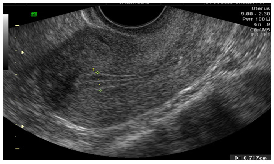
Trans vaginal ultrasound image of a midcycle triple line healthy endometrium with adequate thickness.
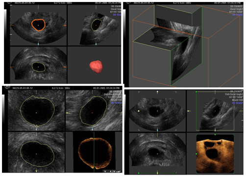
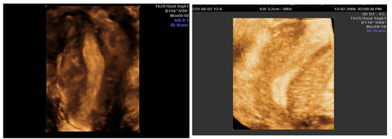
Uterine cavity volume by 3D 4D sonography, a volume of 3 – 5 cc in midcycle phase is optimum for good receptivity.
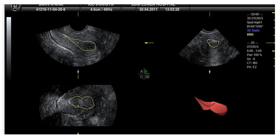
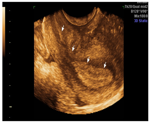
Image of a thickened endometrium (by 3D) as in secretary phase of menstrual cycle.
This image shows follicular volume in pre-ovulatory phase (by 3D – 4D)
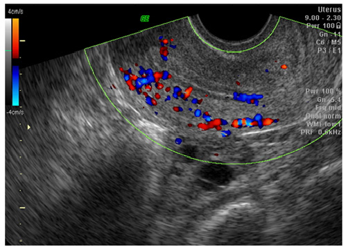
Good Myometrial and endometrial vascularity as seen by colour Doppler in a periovulatory Endometrium.
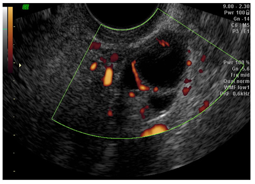
Good perifollicular blood flow as seen by power Doppler.
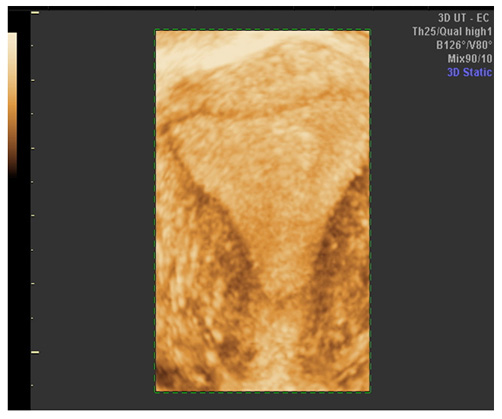
3D image of thickened endometrium.
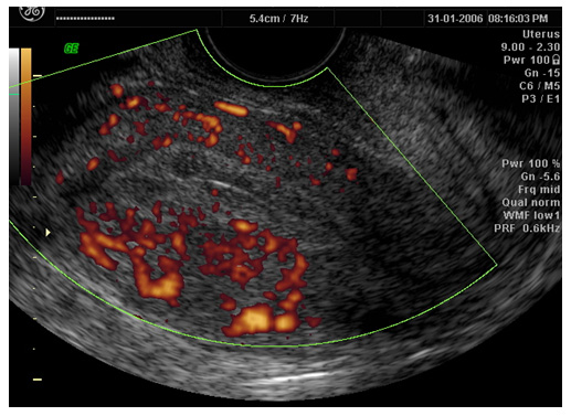
Myometrial blood flow as seen by power doppler internal to the arcuate vessels.
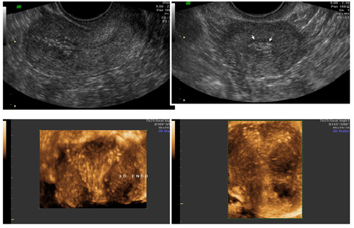
3D and 4D images of a lady with intra uterine adhesions (Asherman syndrome)
Benign endometrial polyps within uterine cavity.
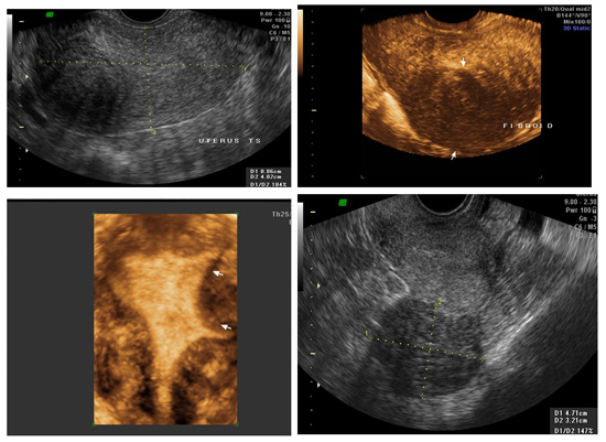
2D and 3D 4D images of different types of fibromyomas of uterus (intramural and subserosal).
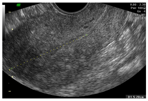
Image of an adenomyotic uterus.
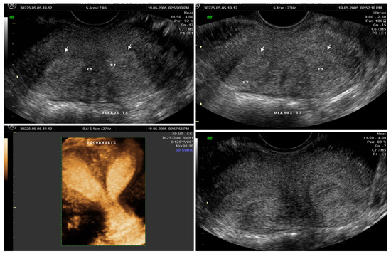
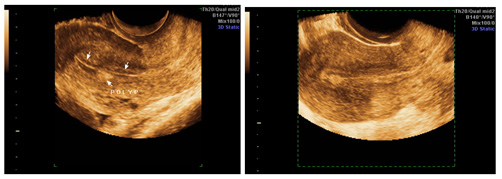
Images show Congenital malformations of uterus – bicornuate uterus.
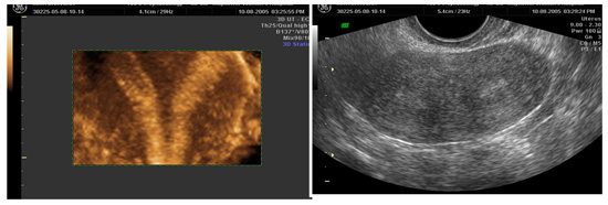
2D and 3D image showing septate uterus.
3D & 4D images of a unicornuate uterus.
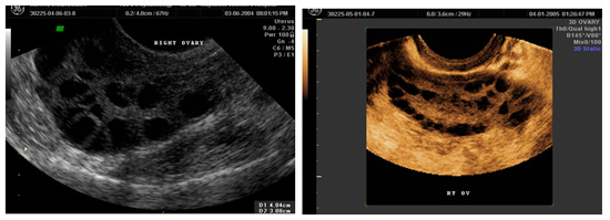
Enlarged polycystic ovaries with multiple small follicles an increased ovarian volume, a common cause of irregular periods and infertility.
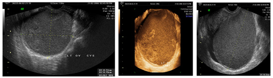
Ovarian endometriomas, the classical chocolate cyst, responsible for dysmenorrhoea and infertility in many women today.